The Royce 3D Bioelectronics Facility at the University of Cambridge is designed for physical scientists and engineers to test the interaction of 3D materials and bioelectronic devices with cells.
Our focus is on facilitating researchers with limited access to cell biology equipment in the fabrication and subsequent analysis of 3D-cell interfacing constructs.
Cell-biological training provided at the 3D Bioelectronics Facility encourages networking between researchers from a range of disciplines. This facility is embedded within the Cambridge Centre for Medical Materials at the Department of Materials Science and Metallurgy to capitalise on their expertise.
For information about using the equipment contact us at royce@maxwell.cam.ac.uk
Equipment falls into six categories:
- Ice-templating equipment to fabricate porous 3D materials from aqueous suspensions.
- Cell culture equipment for growth and maintenance of cells in 3D culture.
- Electrical acquisition equipment to analyse cell behaviour on novel materials and bioelectronic devices.
- Simultaneous optical and electrical measurement of cells and materials via two-photon microscopy with integrated electrophysiology.
- Apparatus to section 3D samples.
- Equipment to transport live cell containing devices to and from the 3D Bioelectronics Facility
A short video tour of the facility is available here.
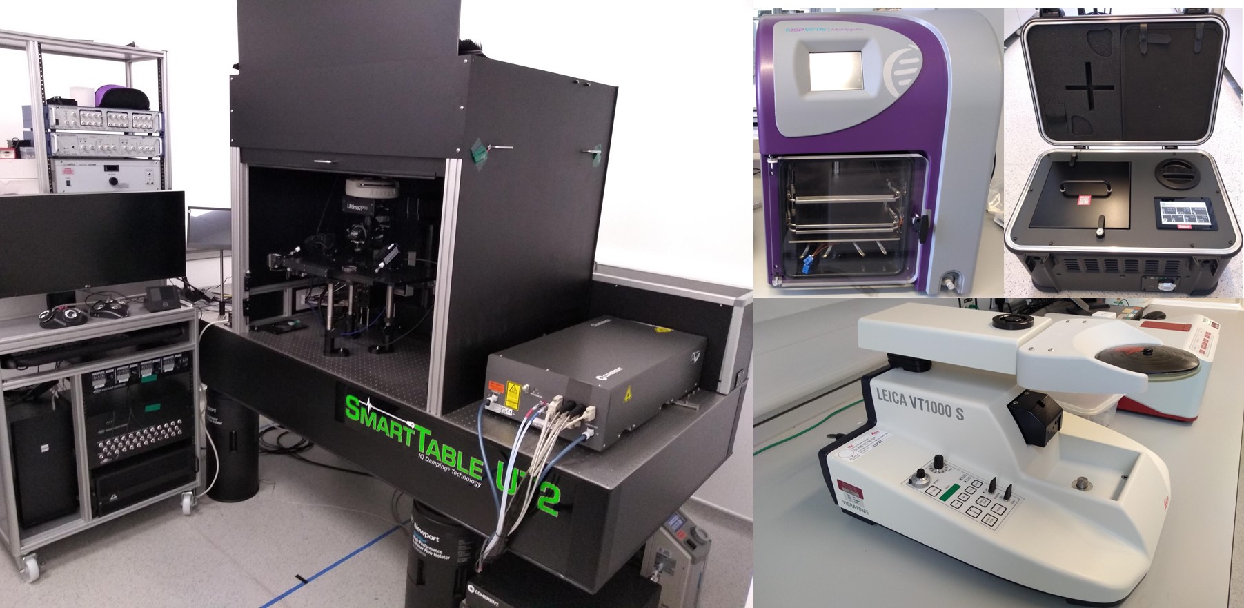
Basic Description: The VirTis AdVantage Pro freeze dryer freezes and lyophilises solutions and suspensions to produce 3D scaffold materials.
Detailed Description: The VirTis AdVantage Pro freeze dryer is used to freeze samples under defined conditions, such as freezing rate and temperature. This carefully controlled freezing forms ice crystals with specific geometry within the solution/suspension that are subsequently removed via sublimation. This is achieved by thawing the sample at low pressure. The solvent evaporates directly from the solid phase to the vapour phase without going through the liquid phase. 3D structures are formed where the pores represent the regions where the solvent crystals were removed.
The VirTis AdVantage Pro freeze dryer has two 273mm x 355mm fluid cooled production shelves with a shelf clearance of 73mm. The shelf temperature can range from -55°C to +60°C and the condenser has a total capacity of 6 litres and minimum temperature of -82°C. Four product probes are incorporated to record the temperature profiles within samples. The user interface is via the Intellitronics™ Touch-Screen Controller with recipe customization of up to 10 thermal treatment and 12 drying steps.
Uses: Typically, the VirTis AdVantage Pro freeze dryer is used to lyophilise macro-molecular solutions and suspensions. This can allow storage of unstable molecules under ambient conditions, or the production of highly porous 3D materials. Porous 3D materials produced by freeze drying are highly suited to biomaterial applications. Their pore dimensions, often in the 40-200μm range, and interconnectivity between pores permits cellular infiltration. Freeze drying lacks the harsh conditions of other 3D material fabrications techniques, so retains the biological activity of the precursor molecules.
Basic Description: A full cell culture laboratory for the routine culture of mammalian cells and preparation, culture, and analysis of cell-loaded scaffolds and bioelectronic devices.
Detailed Description: The full containment level II laboratory for the culture and analysis of mammalian cell includes:
- ThermoFisher 1.2m wide Advantage Containment Level II Microbiological Safety Cabinet with integrated UV light.
- Two sets of PHCbi 165 litre CO2 incubators. One for routine culture and the other for quarantine of new cell lines or installation of electrophysiology rigs. These routinely operate at 5% CO2 and ambient oxygen.
- Refrigerated ThermoFisher 5804R G Centrifuge fitted with a A444 swing bucket rotor and 15/50ml tube adapters.
- A Grant SUB Aqua Pro SAPD Water bath with independent temperature regulation on a 5L and 12L bath.
- EVOS XL Core Mechanical Stage transmitted light inverted phase contrast microscope imaging system. This has an integrated colour camera and tilting LCD. Its four-position turret is configured with 4X, 10X, 20X and 40X phase objectives.
- ThermoFisher COUNTESS 3 automated cell counter.
Uses: Typically, the cell culture lab is used for mammalian cell expansion and seeding cells onto scaffolds, materials, and devices. These include established primary and transformed cell lines. The cell lab can be used for cell analysis. This includes continuous bulk electrophysiological measurement of live cells-loaded experimental devices, colorimetric assays or preparation of samples for immunofluorescence microscopy.
Basic Description: Equipment to take bulk electrophysiological measurements of cell-loaded bioelectronic devices and 3D scaffolds.
Detailed Description: A Metrohm PGSTAT302N potentiostat with FRA32M Module and an Intan RHD USB interface board including RHD 64-channel head-stage with accelerometer. The PGSTAT302N is a high-end, high current potentiostat/galvanostat, with a compliance voltage of 30 V and a bandwidth of 1 MHz. Combined with the FRA32M module, this equipment is specially designed for electrochemical impedance spectroscopy. The maximum current is 2 A and the current resolution is 30 fA at a current range of 10 nA. The Intan RHD USB interface board records electrophysiology signals from 64 channels using small foot-print hardware and free, open-source software. The board connects to a host computer via a USB 2.0 cable and the RHD head-stage connects to the recording controller via thin, flexible all-digital SPI cables.
Uses: The Metrohm PGSTAT302N potentiostat is an essential analytical instrument for studying electrochemical devices and reactions. It controls the potential of one or more working electrodes to generate and measure potentials and currents in electroanalytical experiments. The Intan RHD USB board allows users to record weak electrical signals produced by biological systems. It transforms these bioelectrical signals directly into digital data.
Basic Description: A two-photon microscope to take fluorescent images up to 800μm deep into a material while conducting simultaneous electrophysiology measurements.
Detailed Description: The Bruker Ultima 2Pplus two-photon laser scanner microscope is fitted with two Hamamatsu GaAsP detectors and Coherent Chameleon Vision II laser. The laser can excite at wavelengths from 680 nm to 1080 nm allowing efficient excitation of a wide variety of fluorescent markers, calcium indicators, and long wavelength probes such as mCherry. Emissions are measured simultaneously in two channels using a 565nm dichroic beam splitter coupled with et525/70m-2P and et595/50m-2P bandpass emission filters. The microscope is fitted with a 16X 0.8NA water immersion objective with oversized optics delivering a 1.4mm x 1.4mm field of view. Fast (30fps) imaging can be acquired using a 8kHz resonant galvanometer, in addition to high-resolution imaging using a standard galvanometer.
The Bruker Ultima 2Pplus is fitted with an oversized stage housing two Sensapex four-axis manipulators controlled by a uMp-TSC touch screen unit and four-wheel controller. These connect to a Molecular Devices 700B multiclamp computer-controlled microelectrode current and voltage clamp amplifier for electrophysiology and electrochemistry measurements. The Molecular Devices Digidata 1550B1 low-noise digitizer digitises inputs across a wide range of signals from -10 to +10 V and sampling rate of 500 kHz while the HumSilencer provides 50/60 Hz noise cancellation.
Uses: The Bruker Ultima 2Pplus 2-photon microscope can be used, independently from the electrophysiology detection equipment, to take fluorescent images through samples up to ~800μm thick. Z-stacks with 1μm depth resolution can be acquired without the need for sectioning, allowing imaging and 3D reconstruction of materials and cells. Tuning of infrared excitation wavelengths permits label-free imaging of biological macromolecules and polymeric material with excellent sampling depth and minimal photo-bleaching. Coupled with the electrophysiology equipment, this can be used for fast electrophysiology protocols and simultaneous optical imaging.
Basic Description: The Leica VT1000S cuts soft materials, such as foams, elastomers and plant roots into thin sections.
Detailed Description: The Leica VT1000S is a vibrating blade microvibratome designed to cut soft materials into thin sections. The blade frequency can be altered between 0 and 100Hz. There is fine control over the blade advance speed between 0.025 to 2.5mm/s. Section thickness can be controlled between 1 and 999μm in 1μm increments. Maximum sample dimensions are 33 x 40 x 15mm. A programmable sectioning window reduces unnecessary blade movement either side of the sample, ensuring quick sectioning of even the smallest specimens.
Uses: The Leica VT1000S is used to cut large, soft samples into thin sections. Samples can be sectioned prior to cell seeding, to yield small samples, so maximising the cell density within a sample. Fixed samples can be sectioned post staining and imaging.
Basic Description: The Cell Box Ground 2.0 is a portable cell culture incubator that is suitable for overland transportation of live cell systems.
Detailed Description: The Cell Box Ground 2.0 produces a cell culture incubator environment for overland transport of cells and cell-loaded materials between sites. It contains a rechargeable lithium-ion battery for stable temperature control between 28-38°C for 48 hours at 37°C. CO2 control for up to 72 hours at 5%CO2 is provided by disposable CO2 canisters. Inserts within the incubator chamber enable use with multi-well plates, tissue culture flasks and tubes and other gas-permeable vessels. The temperature and CO2 concentration are constantly logged via the inbuilt data tracking software.
Uses: The Cell Box Ground 2.0 is used for overland, but not air, transportation of live in vitro cell cultures between research sites for up to two days.