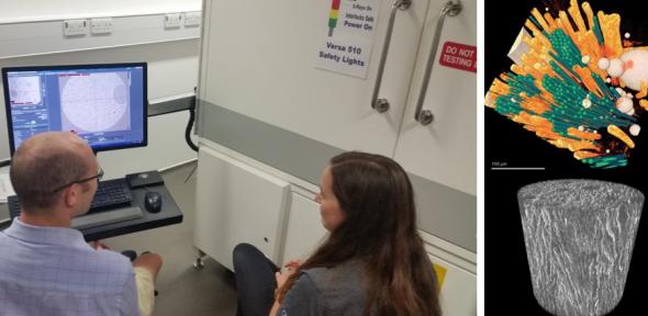
The 3D Computer Tomography (CT) microscope is a ZEISS Versa 510 used for non-destructive, in-situ characterisation and observation of the composition, deformation and damage development of a broad range of materials.
At Cambridge the CT microscope has been used to image a vast range of materials from insects to devices, ceramics, batteries and metals. The system is rarely limited.
Samples can be as small as tens of microns to several inches. The stage can handle up to 15kg, custom stages can be developed.
Our CT microscope can view deeply buried microstructures that may be unobservable with 2D surface imaging. It can also be used for investigating samples with features on length scales from 50 microns down to 1 micron.
It is useful for
- generating an accuarte 3D image
- determining the relationship between processing and microstructure
- quantifying and characterising microstructural evolution
- performing in-situ and 4D (time dependent) studies to understand the impact of heating, cooling, oxidation, wetting, tension, tensile compression, imbibition, drainage and other simulated environmental studies
If your research interests require specialised 3D observation of microstructure evolution and you have any questions on the tool’s capabilities or would like to discuss your experimental needs, contact technical lead Tony Dennis (ad466@cam.ac.uk) in the Department of Engineering.
